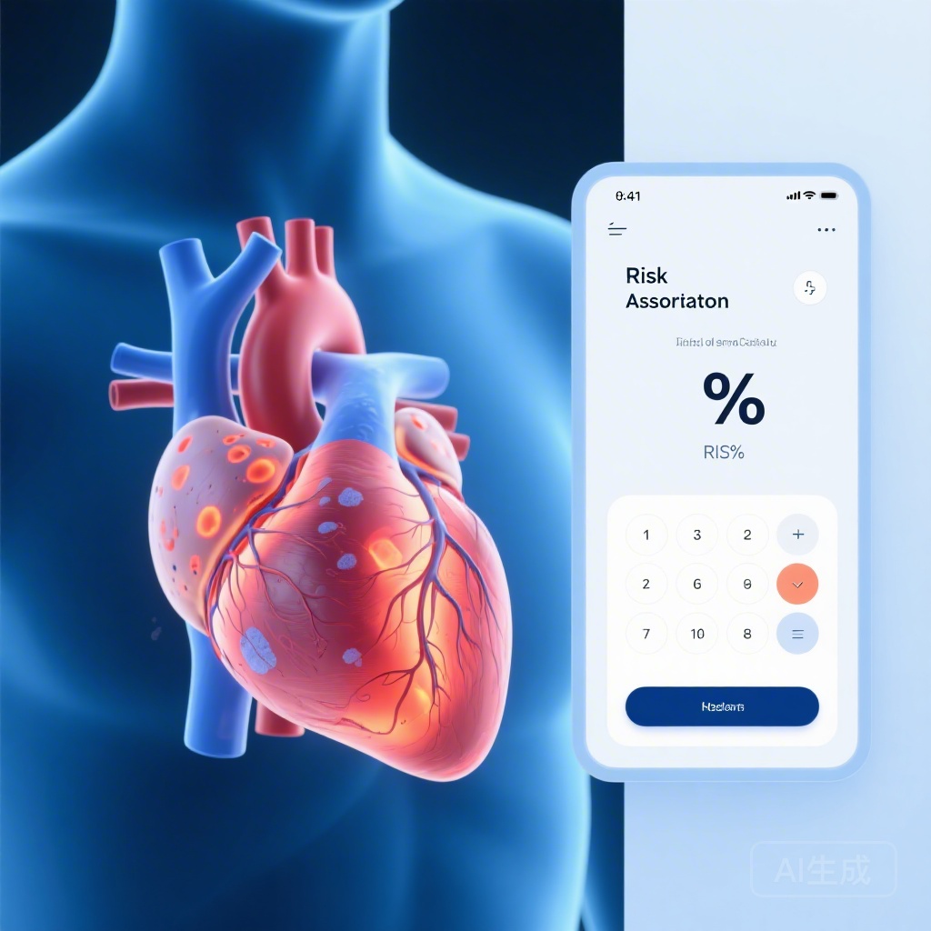
Introduction: The Evolving Landscape of ARVC Risk Assessment
Arrhythmogenic Right Ventricular Cardiomyopathy (ARVC) remains one of the most challenging conditions for clinical electrophysiologists and cardiologists. Characterized by the progressive replacement of myocardium with fibro-fatty tissue, ARVC is a leading cause of sudden cardiac death (SCD) in young adults and athletes. Historically, risk stratification has relied on the 2010 Task Force Criteria (TFC), which focused heavily on right ventricular (RV) morphology and electrical instability. However, the development of the ARVC risk calculator—a validated tool that estimates the risk of incident sustained ventricular arrhythmia (VA)—represented a significant leap forward in personalized medicine.
Despite the success of the ARVC risk calculator, a persistent question in the field has been the role of left ventricular (LV) involvement. As our understanding of ARVC has shifted from a purely right-sided disease to a potentially biventricular or even left-dominant condition, clinicians have increasingly looked toward Cardiac Magnetic Resonance (CMR) imaging, specifically Late Gadolinium Enhancement (LGE), to identify high-risk phenotypes. The study by De Marco et al., published in Circulation: Arrhythmia and Electrophysiology, seeks to answer whether the presence of LV LGE provides a necessary refinement to the existing risk models.
Highlighting Key Insights
1. LV LGE was detected in approximately 34% of the study cohort, suggesting that left ventricular involvement is relatively common even in patients meeting the 2010 Task Force Criteria.
2. While univariable analysis showed a nearly two-fold increase in VA risk for patients with LV LGE, this association vanished when adjusted for the parameters already included in the ARVC risk calculator.
3. The study reinforces that the current ARVC risk calculator is a robust and sufficient tool for predicting first-time sustained ventricular arrhythmias, even without the inclusion of complex CMR-based LV imaging markers.
Study Design and Methodology
This robust investigation was conducted across 17 specialized centers, involving a cohort of 385 patients. To ensure the findings were applicable to primary prevention scenarios, the inclusion criteria were strict: patients must have had a diagnosis of definite ARVC, no prior history of sustained VA, and have undergone contrast-enhanced CMR at baseline.
Patients were followed for a median of 3.1 years. The primary endpoint was the occurrence of a first sustained VA, defined as sustained ventricular tachycardia, ventricular fibrillation, or an appropriate implantable cardioverter-defibrillator (ICD) intervention. The researchers specifically looked for the presence of LV LGE and categorized a subset of patients as having “high-risk” LGE patterns, which included epicardial, transmural, or combined septal and free-wall enhancement. Cox proportional hazard models were employed to determine whether these imaging findings offered incremental prognostic value over the validated ARVC risk calculator score.
Key Findings: The Limits of Imaging in Risk Prediction
The demographic profile of the cohort was typical for ARVC: a mean age of 39.6 years, approximately 40% male, and a high proportion of probands (54%). During the follow-up period, 17.4% (67 patients) reached the primary endpoint of sustained VA.
Initial Associations
At the univariable level, the presence of LV LGE was indeed a red flag. Patients with any LV LGE had a hazard ratio (HR) of 1.82 (P=0.014) for experiencing a VA. Those with high-risk LV LGE patterns showed a similar risk profile (HR, 1.85; P=0.017). These findings initially suggested that CMR imaging could be a powerful tool for identifying those at greatest risk.
The Impact of Multivariate Adjustment
However, the clinical utility of a marker is defined by its incremental value—whether it tells us something we don’t already know. When the researchers adjusted the model for the risk predicted by the standard ARVC risk calculator (which includes variables like age, sex, T-wave inversion on ECG, and RV ejection fraction), the significance of LV LGE disappeared entirely. The P-value for the presence of LV LGE rose to 0.85, and for high-risk LV LGE, it rose to 0.87.
This indicates that the prognostic information provided by LV LGE is likely already captured by the other clinical and electrical markers integrated into the risk calculator. In other words, LV LGE is a marker of more advanced disease, but it does not appear to be an independent driver of arrhythmia risk once other disease manifestations are accounted for.
Expert Commentary: Interpreting the Data
The results of this study offer a sobering but practical message for clinicians. While the presence of LV LGE on a CMR report often causes clinical anxiety and may tempt a physician toward more aggressive interventions like ICD implantation, this data suggests caution.
One possible explanation for these findings is that LV involvement in ARVC typically occurs in tandem with significant RV dysfunction or extensive electrical remodeling (manifesting as T-wave inversions across multiple leads). Since the ARVC risk calculator already incorporates these parameters, the addition of LGE provides redundant information. Furthermore, while LGE represents structural fibrosis, the actual trigger for VA in ARVC is often a complex interplay of genetic predisposition, mechanical strain, and inflammatory signaling that may not be fully captured by a static imaging snapshot of the left ventricle.
It is also important to note the study’s limitations. The median follow-up of 3.1 years, while substantial, may not capture the very long-term risk associated with progressive LV fibrosis. Additionally, as ARVC is a heterogeneous disease, there may still be specific sub-phenotypes—such as those with isolated left-dominant disease—where LGE plays a more prominent role, though such patients were not the primary focus of this TFC-based cohort.
Conclusion: Clinical Takeaways for Practice
The study by De Marco et al. clarifies the role of CMR in the routine management of ARVC. For the prediction of incident sustained ventricular arrhythmia, the validated ARVC risk calculator remains the gold standard. While LV LGE is a useful marker for characterizing the extent of disease and confirming a diagnosis, its presence should not, in isolation, override the risk estimates provided by the clinical calculator.
Clinicians should continue to prioritize the comprehensive data points required by the calculator—including 24-hour Holter monitoring for PVC count, detailed ECG analysis, and RV functional assessment—to make informed decisions regarding ICD implantation. The search for new biomarkers continues, but for now, the established clinical models hold their ground against advanced imaging metrics.
References
De Marco C, Asatryan B, Te Riele ASJM, et al. Left Ventricular Late Gadolinium Enhancement for Arrhythmic Risk Prediction in ARVC. Circ Arrhythm Electrophysiol. 2026 Feb;19(2):e014265. doi: 10.1161/CIRCEP.125.014265. Epub 2026 Jan 29. PMID: 41608798.