Introduction: The Shifting Landscape of Autoimmune Gastritis
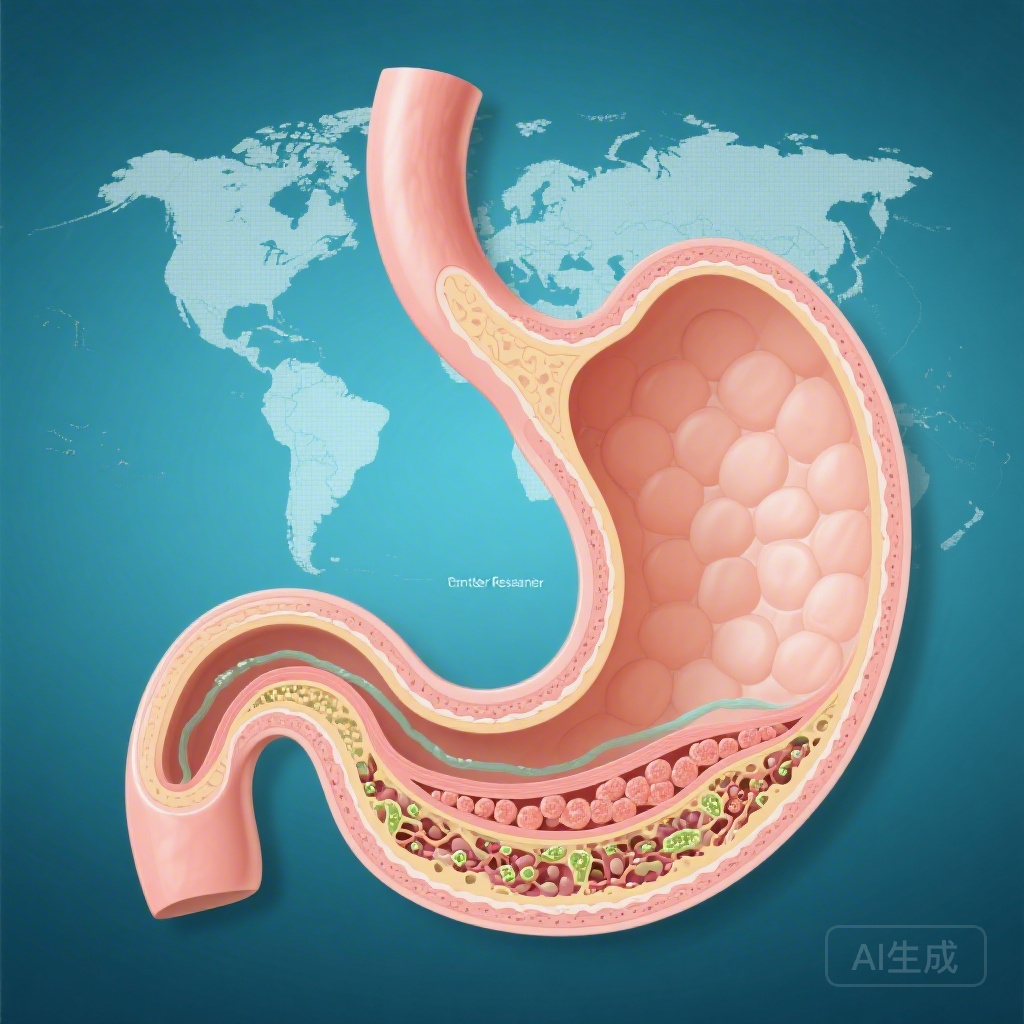
Autoimmune gastritis (AIG) is a chronic, progressive inflammatory condition characterized by the immune-mediated destruction of parietal cells in the gastric corpus and fundus. Historically viewed as a rare cause of pernicious anemia in elderly populations of Northern European descent, our understanding of AIG has undergone a paradigm shift. Recent evidence suggests that AIG is more prevalent and geographically diverse than previously recognized, often presenting in younger cohorts and manifesting through a variety of clinical phenotypes beyond classical vitamin B12 deficiency.
Crucially, the long-term sequelae of AIG include a significantly elevated risk for gastric neoplasia, specifically gastric adenocarcinoma and type 1 gastric neuroendocrine tumors (NETs). However, until recently, large-scale international data comparing the clinical profile and neoplastic outcomes across different ethnic and geographic populations have been scarce. A landmark multicentre study led by Lenti et al., published in Gut, provides essential insights into these regional variations and identifies key clinical predictors for malignant progression.
Highlights of the International Multicentre Study
This research represents one of the most comprehensive efforts to characterize AIG on a global scale. Key highlights include:
1. Clinical heterogeneity is marked by geographic region, with microcytic anemia predominating in Turkish and Latin American cohorts, while macrocytic anemia remains the primary presentation in Europe.
2. The incidence of gastric adenocarcinoma was recorded at 2.9%, with risk significantly increased in patients over 65 years and those with advanced histological changes.
3. Type 1 gastric neuroendocrine tumors (NETs) were observed in over 10% of the cohort, with smoking and proton pump inhibitor (PPI) use identified as notable risk factors.
4. Helicobacter pylori eradication appears to exert a protective effect against the development of neuroendocrine tumors in the context of AIG.
Disease Burden and Clinical Context
AIG remains an underdiagnosed condition, often remaining silent for years before the onset of symptomatic anemia or neoplastic complications. The disease burden is not merely limited to nutritional deficiencies (iron and B12) but extends to the psychological and economic costs of long-term surveillance for gastric cancer. The destruction of parietal cells leads to achlorhydria, which in turn triggers compensatory G-cell hyperplasia and profound hypergastrinemia. This hormonal imbalance is the primary driver for ECL-cell hyperplasia and the subsequent development of type 1 NETs. Furthermore, the chronic inflammatory milieu and the development of intestinal metaplasia create a fertile ground for the Correa cascade, leading to adenocarcinoma.
Study Design and Population Characteristics
This retrospective study included 1,240 adults with histologically confirmed AIG from eight tertiary centers across Europe, Türkiye, Latin America, the USA, and Japan. The cohort was predominantly female (2:1 ratio) with a median age of 59 years. The researchers collected extensive longitudinal data with a median follow-up of 68 months, allowing for a robust estimation of the crude incidence rates of gastric malignancies.
The study was designed to evaluate not only the frequency of neoplastic events but also the diverse clinical presentations. By comparing data across continents, the authors were able to highlight how AIG might be detected earlier in some regions through the identification of microcytic anemia (iron deficiency), which typically precedes the macrocytic anemia (B12 deficiency) characteristic of end-stage gastric atrophy.
Key Findings: Regional Clinical Phenotypes
The study revealed striking differences in how AIG presents clinically across the globe. In Europe, macrocytic anemia was the most common hematological finding (45.6%). In contrast, microcytic anemia was the dominant presentation in Türkiye (56.1%) and Latin America (64.7%). This suggests that in certain populations, the loss of gastric acid (required for iron absorption) is the first clinically detectable sign of parietal cell loss, long before the loss of intrinsic factor leads to B12 malabsorption.
Furthermore, autoimmune comorbidities—such as autoimmune thyroid disease or type 1 diabetes—were most frequent in the Latin American cohort (67.7%). These findings emphasize that clinicians must maintain a high index of suspicion for AIG in patients with unexplained iron deficiency or existing autoimmune disorders, regardless of their age or ethnicity.
Neoplastic Risks: Quantifying Adenocarcinoma and NET Incidence
Over the follow-up period, 36 cases of gastric adenocarcinoma (2.9%) and 132 cases of NETs (10.6%) were identified. The crude incidence rates ranged from 1.15 to 1.47 per 100 person-years for adenocarcinoma and 0.70 to 1.62 per 100 person-years for NETs. Interestingly, no incident adenocarcinomas were reported in the Latin American or Japanese cohorts during the study period, although this may be influenced by local screening practices or genetic factors.
Predictors of Gastric Adenocarcinoma
The study identified several critical factors associated with the development of adenocarcinoma:
1. Age >65 years: Patients in this age group had a 4.5-fold increase in risk (OR 4.50, 95% CI 2.18 to 9.27).
2. Intestinal Metaplasia: The presence of intestinal metaplasia on histology was a significant predictor (OR 1.51, 95% CI 1.16 to 1.97).
3. Extreme Hypergastrinemia: Gastrin-17 levels exceeding 1316 pg/mL were associated with a staggering 15-fold increase in risk (OR 15.52, 95% CI 3.61 to 66.71).
4. Prior PPI Use: History of PPI use was associated with higher odds (OR 5.74, 95% CI 2.13 to 15.47), likely reflecting a diagnostic delay where PPIs were prescribed for dyspeptic symptoms before AIG was identified.
Risk Factors for Type 1 Gastric Neuroendocrine Tumors
For NETs, the risk profile was slightly different:
1. Smoking: Current or former smokers had a 2.45-fold higher risk.
2. Intestinal Metaplasia: Similar to adenocarcinoma, IM increased the odds of NETs (OR 2.88).
3. Gastrin-17: High levels (>1316 pg/mL) were again a strong predictor (OR 3.25).
4. H. pylori Eradication: Notably, patients who underwent successful H. pylori eradication had significantly lower odds of developing NETs (OR 0.25, 95% CI 0.07 to 0.88), suggesting that treating the infection may mitigate the hypergastrinemic drive.
Expert Commentary: Mechanistic Insights and Clinical Implications
The association between high Gastrin-17 levels and adenocarcinoma is particularly provocative. While hypergastrinemia is well-known to drive NETs through ECL-cell stimulation, its role in adenocarcinoma may be more complex, potentially involving the stimulation of cholecystokinin-2 (CCK2) receptors on other gastric cells or simply serving as a high-fidelity biomarker for the severity of corpus atrophy.
The finding regarding PPI use warrants careful interpretation. It is unlikely that PPIs cause gastric cancer in these patients; rather, it is more probable that patients with early, undiagnosed AIG present with vague dyspepsia and are empirically treated with PPIs. This highlights the danger of ‘symptomatic’ treatment without a definitive diagnosis in patients with refractory dyspepsia or anemia.
From a clinical standpoint, the protective effect of H. pylori eradication against NETs is a vital takeaway. It suggests that even in the presence of established autoimmune atrophy, removing the synergistic inflammatory stimulus of H. pylori can alter the neoplastic trajectory. This supports a policy of ‘search and treat’ for H. pylori in all patients diagnosed with AIG.
Conclusion: Moving Toward Precision Surveillance
The Lenti et al. study underscores that autoimmune gastritis is a global disease with regional nuances. The significant differences in anemia patterns and neoplastic rates suggest that a ‘one-size-fits-all’ approach to follow-up may be suboptimal. Instead, surveillance strategies should be tailored based on geographic risk profiles, age, and specific biomarkers like Gastrin-17.
For the practicing clinician, the message is clear: evaluate iron deficiency as a potential early marker of AIG, perform high-quality endoscopy with mapping biopsies in suspected cases, and utilize Gastrin-17 and histological findings (specifically intestinal metaplasia) to risk-stratify patients for more intensive cancer surveillance. Future research should focus on prospective validation of these risk factors and the development of cost-effective screening protocols in non-European populations.
References
1. Lenti MV, Miceli E, Soykan I, et al. Novel insights into autoimmune gastritis: clinical profile and gastric neoplastic risk from an international multicentre study. Gut. 2026;75(3). PMID: 41791849.
2. Rugge M, Lenti MV, Giuffrida P, et al. Autoimmune gastritis: A biopsy-based pathology. American Journal of Surgical Pathology. 2020;44(12):1564-1574.
3. Neumann WL, Coss E, Rugge M, Genta RM. Autoimmune gastritis. Autoimmunity Reviews. 2013;12(5):554-560.
4. Shah SC, Piazuelo MB, Kuipers EJ, Li D. AGA Clinical Practice Update on the Diagnosis and Management of Atrophic Gastritis: Expert Review. Gastroenterology. 2021;161(4):1325-1332.