Highlight
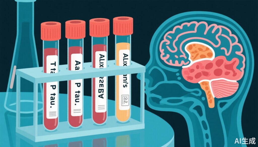
- Three core cerebrospinal fluid biomarkers—T-tau, P-tau, and Aβ42—show strong diagnostic accuracy for Alzheimer’s disease.
- Emerging markers such as CSF neurofilament light (NFL) and plasma T-tau also demonstrate significant association with Alzheimer’s disease.
- Several other CSF biomarkers moderately relate to Alzheimer’s pathology, while plasma Aβ42 and Aβ40 lack significant diagnostic value.
- Recommendations support routine use of core CSF biomarkers in both clinical diagnosis and research settings.
Study Background
Alzheimer’s disease (AD) remains the leading cause of dementia worldwide, imposing substantial clinical, social, and economic burdens due to its progressive neurodegenerative nature. Early and accurate diagnosis is crucial for appropriate management and future therapeutic intervention trials. Biomarkers obtained from cerebrospinal fluid (CSF) and blood provide biological evidence of underlying AD pathology, facilitating early detection before overt cognitive decline. Over decades, core CSF biomarkers—comprising amyloid-beta peptide 42 (Aβ42), total tau protein (T-tau), and phosphorylated tau (P-tau)—have become central in AD research and diagnosis. In addition, emerging biomarkers reflecting neuronal injury, glial activation, and blood-brain barrier dysfunction hold promise to improve diagnostic precision. Despite numerous original studies, there has been a lack of comprehensive quantitative synthesis regarding the diagnostic performance of these markers across diverse cohorts. This systematic review and meta-analysis addresses this gap by evaluating 15 candidate biomarkers in CSF and blood, assessing their utility in differentiating AD from controls and mild cognitive impairment (MCI) subtypes.
Study Design
This meta-analysis systematically identified relevant studies published between July 1, 1984, and June 30, 2014, by searching PubMed and Web of Science databases. Included studies featured cohorts with Alzheimer’s disease and appropriate control groups, including cognitively normal individuals and patients with stable mild cognitive impairment. Fifteen biomarkers were investigated, categorized mechanistically as follows:
- Neurodegeneration markers: T-tau, neurofilament light chain (NFL), neuron-specific enolase (NSE), visinin-like protein 1 (VLP-1), heart-type fatty acid binding protein (HFABP)
- Amyloid precursor protein (APP) metabolism: Aβ42, Aβ40, Aβ38, soluble APP alpha (sAPPα), and soluble APP beta (sAPPβ)
- Tangle pathology: Phosphorylated tau (P-tau)
- Blood-brain barrier integrity: Albumin ratio
- Glial activation: YKL-40, monocyte chemoattractant protein-1 (MCP-1), and glial fibrillary acidic protein (GFAP)
Data extraction and quality assessment were performed by multiple authors using a modified QUADAS tool. Biomarker performance was analyzed using random-effects meta-analyses calculating the ratio of biomarker concentrations between AD and control groups, and between MCI due to AD and stable MCI cohorts with at least two years follow-up.
Key Findings
A total of 231 articles encompassing 15,699 AD patients and 13,018 controls were included. The core CSF biomarkers presented robust differentiating power:
- T-tau: average ratio 2.54 (95% CI 2.44–2.64; p<0.0001)
- P-tau: 1.88 (95% CI 1.79–1.97; p<0.0001)
- Aβ42: 0.56 (95% CI 0.55–0.58; p<0.0001) indicating reduced levels in AD
These biomarkers also distinguished MCI due to AD from stable MCI effectively:
- CSF Aβ42 ratio 0.67
- P-tau ratio 1.72
- T-tau ratio 1.76
Beyond the core triad, additional biomarkers showed notable diagnostic value:
- CSF NFL had a large effect size (2.35, 95% CI 1.90–2.91; p<0.0001), reflecting axonal damage in AD.
- Plasma T-tau also differentiated AD from controls with a significant effect (1.95, 95% CI 1.12–3.38; p=0.02), supporting potential for less invasive diagnostics.
- Moderate effect sizes were observed for CSF NSE, VLP-1, HFABP, and YKL-40 (ratios ranging 1.28–1.47), markers associated with neuronal injury and glial responses.
- Other biomarkers, including plasma Aβ40 and Aβ42, albumin ratio, MCP-1, and GFAP, demonstrated minimal or no significant changes between groups.
The overall findings underscore the stability and consistency of core CSF biomarkers in AD diagnosis, supported by independent validation across large populations and heterogeneous cohorts.
Expert Commentary
This comprehensive meta-analysis consolidates substantial evidence supporting established core CSF biomarkers—T-tau, P-tau, and Aβ42—as reliable biological indicators of AD pathology, suitable for widespread clinical use. The strong diagnostic performance in both dementia-stage AD and prodromal MCI due to AD confirms their utility for early detection and patient stratification in trials. The compelling signal from CSF NFL further augments the biomarker repertoire by providing a marker highly sensitive to neuroaxonal damage. Plasma T-tau’s significant albeit moderate effect size encourages continued development of blood-based assays, which could transform screening and monitoring approaches due to their accessibility and reduced invasiveness.
Moderate associations observed for markers such as NSE and YKL-40 highlight the added value of including biomarkers reflecting neuronal damage and neuroinflammation. However, variability in assay methods and limited longitudinal data for some emerging markers temper enthusiasm for immediate clinical adoption. Additionally, plasma amyloid peptides did not perform well, likely reflecting peripheral metabolism and clearance confounding their diagnostic specificity.
Study strengths include rigorous inclusion criteria, broad biomarker coverage, and sophisticated meta-analytic methods yielding clinically meaningful effect sizes. Limitations relate to heterogeneity in assay platforms, study populations, and diagnostic criteria over the three-decade literature span, potentially influencing effect size estimates. Standardization initiatives and harmonized protocols are essential to further refine biomarker thresholds and validate emerging candidates. Future research should integrate multimodal biomarker panels encompassing CSF, blood, and imaging to enhance diagnostic accuracy and disease staging.
Conclusion
This meta-analysis provides compelling evidence endorsing the core CSF biomarkers T-tau, P-tau, and Aβ42 as gold standards for Alzheimer’s disease diagnosis in clinical and research settings. CSF neurofilament light and plasma T-tau emerge as promising complementary markers of neurodegeneration. Other biomarkers such as NSE, VLP-1, HFABP, and YKL-40 may have adjunctive roles but require further validation. Plasma Aβ species do not currently offer reliable diagnostic value. Embracing these robust biomarkers can improve early detection, differential diagnosis, and patient selection for therapeutic trials, ultimately facilitating personalized approaches to Alzheimer’s disease management. Ongoing efforts should focus on assay standardization, integration of blood-based markers, and longitudinal validation to translate these insights into routine practice effectively.
Funding and Disclosures
This work was supported by the Swedish Research Council, Swedish State Support for Clinical Research, Alzheimer’s Association, Knut and Alice Wallenberg Foundation, Torsten Söderberg Foundation, Alzheimer Foundation (Sweden), European Research Council, and Biomedical Research Forum.
References
Olsson B, Lautner R, Andreasson U, Öhrfelt A, Portelius E, Bjerke M, et al. CSF and blood biomarkers for the diagnosis of Alzheimer’s disease: a systematic review and meta-analysis. Lancet Neurol. 2016 Jun;15(7):673-684. doi: 10.1016/S1474-4422(16)00070-3. Epub 2016 Apr 8. PMID: 27068280.