Introduction: An Enigmatic Pain That Defies Amputation
Phantom limb pain is a perplexing condition experienced by many amputees where they continue to sense, often painfully, the presence of a limb that no longer exists. Despite advances in surgical techniques and rehabilitation, this phenomenon has long baffled scientists and clinicians alike. Recently, groundbreaking brain imaging research published in Nature Neuroscience has uncovered pivotal clues explaining why these phantom pains persist, potentially transforming approaches to treatment and neuroprosthetic development.
The Brain’s Body Map: Understanding the Somatosensory Cortex
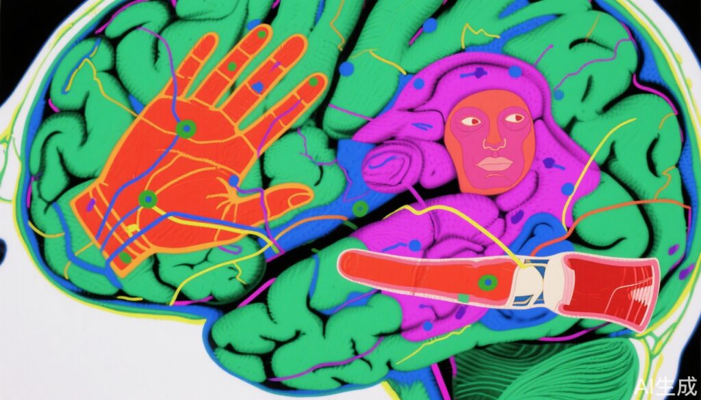
Our brain contains a remarkable feature called the somatosensory cortex, which acts like a neural “body map.” This map has distinct regions corresponding to different body parts, responsible for processing sensations such as touch, temperature, pain, and proprioception (our sense of body position). For example, the region activated when you touch something hot with your hand differs from the area engaged when you stub your toe.
A longstanding question is how this map changes when part of the body is removed, such as an arm or leg amputation. For over five decades, many researchers believed that the brain undergoes significant reorganization: surrounding regions on the map would “take over” the area previously representing the missing limb. This cortical plasticity theory was largely based on observations made after amputation.
However, these assumptions lacked direct comparison with the state of the body map before amputation. Moreover, many amputees report persistent sensations—sometimes accompanied by itching or pain—of their missing limb, challenging the idea that the brain simply rewires itself and erases the lost body part’s representation.
Reevaluating Phantom Limb Phenomena With New Imaging Techniques
Recent brain imaging studies using functional magnetic resonance imaging (fMRI) have shown that when amputees attempt to “move” fingers on their phantom hand, their brain activity patterns resemble those of healthy individuals. This discovery raised essential questions about the nature and stability of the somatosensory cortex after limb loss.
The debate remained unresolved: Does amputation trigger large-scale cortical reorganization, or does the brain’s body map remain more stable than previously thought?
The Landmark Study: Tracking Brain Maps Before and After Amputation
A novel study conducted by researchers at the University of Cambridge has profoundly advanced our understanding. For the first time, they tracked three adult participants before and up to five years after arm amputation using fMRI, mapping their hand and face (specifically lips) representations in the cortex.
Before amputation, participants performed movements including finger taps, lip pursing, and toe bending while their brain activity was recorded to delineate the cortical maps. Notably, the lip and hand areas are adjacent on the brain’s sensory map.
Following amputation, the participants underwent similar testing at 3 and 6 months post-surgery, imagining moving their lost fingers to activate corresponding brain regions. Some even repeated the tests 18 months and five years after amputation.
Astonishing Results: The Body Map Endures
Comparing the data longitudinally, researchers discovered that the cortical representation of the hand remained remarkably consistent before and after amputation, despite the physical loss of the limb. Contrary to the prevailing belief, the adjacent lip area did not invade the hand region; both maps remained stable and distinct.
To validate the findings, the team examined 26 individuals who had upper limb amputations an average of 23.5 years prior. The brain maps of their hands and lips similarly showed enduring stability.
Clinical Implications: Rethinking Phantom Limb Pain Treatment
This landmark finding challenges the dogma that phantom limb pain arises from maladaptive cortical reorganization. Instead, it suggests that the brain’s body map remains intact, and the root issues may lie elsewhere.
The researchers propose that the true problem arises within the residual limb, where severed nerves lose their normal sensory inputs. Without proper targets, nerves can proliferate abnormally, forming neuromas or sending erratic “noisy” signals to the brain that manifest as pain.
Consequently, treatments solely aiming to “restore” brain maps have shown limited efficacy in randomized controlled trials. More promising approaches involve surgical interventions at the residual limb, such as nerve transfers that reroute nerves to new muscle or skin sites, providing healthier targets and reducing pain. Indeed, among the study’s three participants, one who underwent a complex nerve transfer before amputation experienced significant pain relief, while the other two receiving standard care continued to suffer phantom pains.
Advancing Neuroprosthetics: Stability Means Opportunity
The discovery of enduring cortical body maps after limb loss has profound implications for brain-machine interfaces and neuroprosthetics—technologies designed to restore movement and sensation to amputees.
If the brain’s hand representation remained heavily altered, interfacing with neuroprosthetic devices would face serious hurdles. Stability in these maps means the brain’s motor and sensory areas can still be harnessed to control artificial limbs or provide sensory feedback.
Researchers envision developing interfaces that decode fine motor intentions from the preserved hand area, potentially distinguishing individual finger movements and restoring detailed sensory experiences like texture, shape, or temperature.
Expert Perspectives and Future Directions
Dr. Tamar Makin, lead author of the study from the University of Cambridge, emphasizes, “Our findings open a crucial window for designing therapies addressing phantom limb pain and neuroprosthetic control by recognizing the brain’s preserved body map. This challenges decades of assumptions and encourages a shift towards peripheral nerve interventions and sophisticated brain-machine technologies.”
Future research will aim to refine the understanding of micro-level body representations and integrate this knowledge into clinical and technological advances.
Patient Scenario: John’s Journey with Phantom Limb Pain
John, a 45-year-old engineer, lost his right arm in a workplace accident five years ago. Despite physical rehabilitation and use of a prosthetic arm, he frequently experiences distressing phantom limb pain described as burning and cramping sensations originating from his missing fingers.
Traditional therapies aimed at “rewiring” his brain provided minimal relief. However, following involvement in a trial that included nerve-targeting surgery and advanced neuroprosthetic training, John reports dramatic improvements in pain management and is able to control his robotic hand with surprising dexterity.
John’s case exemplifies the promise highlighted by the new research: enduring brain maps combined with targeted peripheral interventions can improve quality of life for amputees.
Conclusion
This pioneering study reveals that the brain preserves its sensory and motor representation of a lost limb long after amputation. This challenges entrenched theories of cortical reorganization and offers promising avenues for treating phantom limb pain through peripheral nerve management. Furthermore, it supports the feasibility of developing advanced neuroprosthetic devices controlled by stable brain maps.
The findings underscore the intricate relationship between peripheral nerve health and central nervous system structure, calling for integrated approaches in clinical care and technological innovation. As research progresses, amputees like John may look forward to more effective pain relief and enhanced prosthetic integration, restoring function and improving lives.
References
1. Schone H-R, et al. “Gone but not forgotten: brain’s map of the body remains unchanged after amputation.” Nature Neuroscience. 2025.
2. Cambridge University Research News. “Gone but not forgotten: Brain’s map of the body remains unchanged after amputation.” https://www.cam.ac.uk/research/news/gone-but-not-forgotten-brains-map-of-the-body-remains-unchanged-after-amputation
3. Callaway E. “Phantom limb study challenges long-held brain plasticity theories.” Nature. 2025; doi:10.1038/d41586-025-02686-5
4. Makin TR, et al. “Stability of somatosensory maps post-amputation.” Nature Neuroscience 2025; DOI: 10.1038/s41593-025-02037-7