Highlight
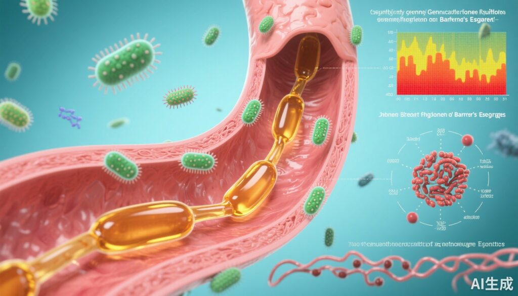
– Elevated secondary bile acids in refluxate are associated with distinct shifts in the esophageal microbiome in Barrett’s esophagus (BE).
– The microbiome changes correlate with significant transcriptomic alterations, including pathways linked to inflammation and oxidative phosphorylation.
– Two distinct gene expression clusters in BE were identified, independent of histologic grade, bile acid concentration, or microbiome composition, suggesting novel molecular subtypes.
– These findings underline the complex interplay between bile acid reflux, microbial ecology, and mucosal gene regulation that may contribute to esophageal adenocarcinoma (EAC) pathogenesis.
Study Background and Disease Burden
Barrett’s esophagus (BE) is a premalignant condition characterized by metaplastic transformation of the esophageal squamous epithelium into specialized intestinal-type mucosa, secondary to chronic gastro-esophageal reflux disease (GERD). BE significantly increases the risk of developing esophageal adenocarcinoma (EAC), a cancer with rising incidence and poor prognosis. The pathogenesis of BE and progression to EAC is multifactorial, involving chronic injury, mucosal inflammation, genetic and epigenetic changes, and more recently recognized, alterations in the esophageal microbiome. Bile acids, a major component of refluxate, especially secondary bile acids formed by bacterial metabolism, are implicated in mucosal injury, inflammation, and carcinogenesis. However, the interactions between reflux bile acids, local microbial communities, and mucosal gene expression in BE remain incompletely understood.
Study Design
This multi-center cross-sectional study enrolled 153 patients undergoing upper endoscopy, including 52 controls with no BE and 101 patients with BE across a spectrum of histopathology: 50 without dysplasia, 10 indefinite for dysplasia, 17 low-grade dysplasia, 17 high-grade dysplasia, and 7 with confirmed EAC. Gastric aspirates were collected to quantify bile acid composition using liquid chromatography–mass spectrometry (LC-MS). Esophageal or gastric cardia tissue biopsies were subjected to 16S rRNA gene sequencing to profile the tissue-associated microbiome, and RNA sequencing was performed on the same tissues to characterize transcriptomic changes. The study compared microbiome composition and gene expression profiles relative to bile acid profiles and histopathologic categories.
Key Findings
The study delineated important associations among refluxate bile acids, esophageal microbiome shifts, and gene expression alterations:
1. Bile Acid Composition: Refluxate samples predominantly contained conjugated bile acids, indicative of minimal microbial deconjugation at the refluxate source. Notably, BE patients exhibited elevated secondary bile acid levels, which are known to be more cytotoxic and pro-inflammatory.
2. Microbiome Alterations: Compared to controls, BE tissues showed a significant increase in Streptococcus species abundance. As dysplasia progressed and in EAC cases, there was an increased prevalence of Lactobacillus and a reduction in Actinomyces and several other genera. These shifts suggest a dynamic microbiome remodeling associated with disease severity.
3. Transcriptomic Correlations: Abundance of Streptococcus correlated strongly with upregulated expression of inflammatory and growth factors such as IL6, FGF2, and HGF. Meanwhile, decreased Actinomyces was linked to widespread gene expression changes including those in the oxidative phosphorylation pathway, highlighting possible metabolic and inflammatory alterations.
4. Gene Expression Clusters: Unsupervised clustering identified two distinct BE gene expression patterns that did not correspond neatly with histologic grade, bile acid profiles, or microbiome composition. This suggests molecular heterogeneity within BE that transcends conventional classification based on dysplasia.
Together, these findings underscore the potential pathogenic role of secondary bile acids in modulating the local microbial environment and tissue gene expression, which may drive carcinogenic pathways in BE.
Expert Commentary
This study by Tanes et al. offers a comprehensive, multi-omic perspective on the triad of reflux bile acids, microbial dysbiosis, and mucosal transcriptome changes in Barrett’s esophagus. The integration of metabolomic, microbiome, and transcriptomic data from well-phenotyped patient samples is a notable strength. The predominance of conjugated bile acids supports clinical observations that bacterial bile acid metabolism occurs largely distal to the stomach, yet elevated secondary bile acids in BE imply localized microbial contributions or altered reflux composition.
The enrichment of Streptococcus and Lactobacillus species with disease progression aligns with prior reports implicating oral and gastric commensals in esophageal pathology, but the specific associations with gene expression of inflammatory mediators enhance mechanistic insight. IL6, FGF2, and HGF are established players in inflammation-driven carcinogenesis and tissue remodeling, and their correlation with specific microbiome shifts presents a plausible causal pathway.
The identification of two molecular clusters independent of histology suggests that future BE risk stratification could incorporate molecular profiling, potentially improving early detection of progression to EAC. However, the cross-sectional design limits causal inference, and longitudinal validation is needed. The absence of extensive bacterial bile acid metabolism in refluxate also prompts further investigation into microbial activities within the esophageal mucosa itself.
Conclusion
This study compellingly demonstrates that increased secondary bile acids in refluxate associate with significant shifts in the esophageal microbiome and mucosal transcriptome in Barrett’s esophagus. These complex interactions may contribute to inflammation, mucosal injury, and carcinogenesis. Understanding these pathways offers opportunities for novel biomarkers and therapeutic targets to improve surveillance and intervene in early neoplastic transformation in BE. Future longitudinal and interventional studies are warranted to explore causality and clinical utility.
References
1. Tanes C, Li Y, Falk GW, Ginsberg GG, Wang KK, Iyer PG, Lightdale CJ, Del Portillo A, Lagana SM, Wang TC, Rustgi AK, Quante M, Jin Z, Wu GD, Friedman ES, Bittinger K, Li H, Abrams JA. Increased reflux secondary bile acids are associated with changes to the microbiome and transcriptome in Barrett’s esophagus. Gut Microbes. 2025 Dec;17(1):2545420. doi: 10.1080/19490976.2025.2545420. Epub 2025 Aug 22. PMID: 40844321; PMCID: PMC12377100.
2. Souza RF, et al. Esophageal Adenocarcinoma: Pathogenesis, Diagnosis, and Treatment. Gastroenterology. 2022;162(3):748-764.
3. Blackett KL, Thomas MG, Girgis S, et al. Evaluation of microbial community profiles in patients with Barrett’s esophagus and esophageal adenocarcinoma. Dig Dis Sci. 2020;65(3):596-606.
4. Bernstein C, Bernstein H, Payne CM, Dvorakova K, Garewal H. Bile acids as carcinogens in human gastrointestinal cancers. Mutat Res. 2005;589(1):47-65.