Study Background and Disease Burden
Wound healing is a complex biological process that plays a crucial role in medical care, particularly in dermatology and surgery. Delays or impairments in wound healing can lead to significant morbidity, prolonged hospitalization, and increased healthcare costs. Chronic wounds, such as diabetic ulcers and pressure sores, represent a substantial burden on patients and health systems, reflecting the necessity for improved evaluation and management strategies. Traditional methods of assessing wound healing often lack precision and repeatability, highlighting a crucial need for advanced techniques that provide detailed insights into the dynamic physiological changes occurring during the healing process.
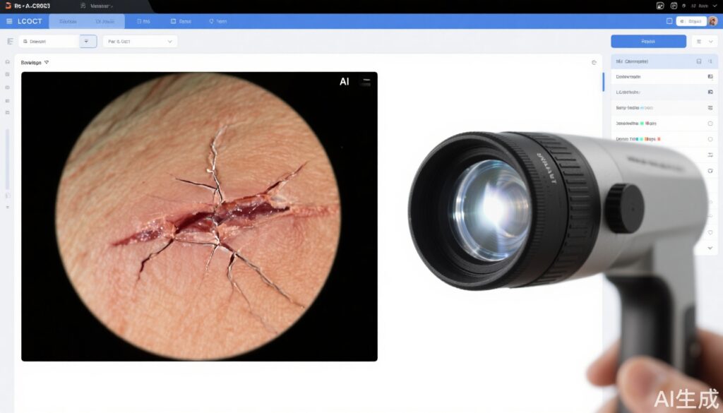
In recent years, ablative fractional photothermolysis has emerged as a promising in vivo model to investigate wound healing. It induces controlled skin injuries that can be monitored using innovative imaging modalities. Line-field confocal optical coherence tomography (LC-OCT) represents a shift towards non-invasive imaging methods, allowing real-time evaluation of skin architecture and function without the need for biopsies or other invasive procedures.
Autonomous analysis through artificial intelligence (AI) algorithms further adds value to this imaging technique, enabling high-throughput evaluation and detailed characterization of wound healing responses at the microscopic level. The fusion of LC-OCT with AI could potentially revolutionize our understanding of wound dynamics and facilitate timely modifications in clinical interventions, ultimately improving patient outcomes.
Study Design
This prospective, randomized, single-blinded pilot study sought to evaluate the combination of LC-OCT and AI algorithms for analyzing acute skin wound healing. Conducted under ethical guidelines, participants were volunteers who underwent fractional ablative CO2 laser treatment at their forearms, with parameters set at 7.5 mJ/MTZ (ClinicalTrials.gov identifier: NCT05614557).
Participants were assigned to three groups: 1) a control group receiving no treatment, and 2) two groups receiving different accredited silicone-based formulations applied to their respective sites. LC-OCT imaging was conducted at preset intervals (from 1 to 7 days) post-laser treatment to capture the temporal and spatial dynamics of wound healing. Subsequently, images were analyzed using proprietary AI algorithms designed to identify and characterize the layered anatomy and biological features associated with skin repair.
Key Findings
The findings showcased the efficacy of LC-OCT in visualizing and quantifying the structural changes in the skin during the wound healing process. Significant differences in healing patterns were detected across the test sites, with the silicone formulations demonstrating superior wound closure rates associated with enhanced structural integrity.
AI algorithms significantly complemented LC-OCT by providing interpretable, quantifiable data on cellular behaviors, particularly keratinocyte migration and proliferation. Parameters such as re-epithelialization rates, collagen deposition, and vascularization could be tracked more effectively through the integrated analysis of imaging data, exceeding what could be achieved using traditional methods.
In terms of clinical implications, primary care physicians and dermatologists are empowered with more robust tools for monitoring treatment efficacy. AI’s capability to delineate skin features allows for immediate adjustments to treatment plans based on individual patient responses, catering to variations in healing trajectories.
Expert Commentary
The amalgamation of LC-OCT with AI is a noteworthy advancement that addresses existing gaps in wound care management. Opinion leaders in dermatology advocate for the implementation of such technologies in routine clinical practice, underscoring their potential to reduce misdiagnosis and provide tailored interventions in wound treatment.
However, the study acknowledges limitations, including the relatively small sample size and the single-center design, which may hinder the generalizability of findings. Future research is needed to validate these results across more diverse populations and to explore long-term outcomes associated with these technological innovations.
Further mechanistic insights into how AI analyzes keratinocyte behavior can deepen our understanding of the biology of skin regeneration. Enhanced biological plausibility emerges when we consider AI’s ability to analyze large datasets, optimizing strategies for wound healing and enabling personalized medicine approaches.
Conclusion
The integration of line-field confocal optical coherence tomography and artificial intelligence heralds a new era in the study of wound healing. This study provides compelling evidence of improved analytical capabilities that offer clinicians valuable insights into the efficacy of wound care interventions. As we advance our understanding of wound healing processes through high-resolution imaging and intelligent algorithms, a path toward refined therapeutic strategies and enhanced patient outcomes comes into focus. Bridging existing research gaps requires ongoing investigation and commitment to implementing innovative technologies across clinical settings.