Highlight
Methotrexate-induced osteopathy is a rare complication seen mainly in postmenopausal women with rheumatoid or psoriatic arthritis. It predominantly manifests as atraumatic stress fractures localized to the lower limbs, particularly the tibia and foot. Early and precise diagnosis using MRI or bone scintigraphy improves clinical identification. Discontinuation of methotrexate significantly enhances patient outcomes, highlighting the need for careful clinical vigilance despite methotrexate’s established therapeutic benefits.
Study Background
Methotrexate (MTX) is a cornerstone drug in managing inflammatory rheumatic diseases such as rheumatoid arthritis (RA) and psoriatic arthritis (PsA). While generally considered safe, rare adverse effects like methotrexate-induced osteopathy have emerged as clinically significant, particularly as these patients often face concurrent osteoporosis risks. Methotrexate-induced osteopathy describes a condition of compromised bone integrity marked by atraumatic stress fractures primarily in the lower extremities. This complication may easily be misdiagnosed or delayed in recognition, given overlapping symptomatology with arthritic joint pain and common osteoporotic fractures. Thus, understanding the patterns, risk factors, and outcomes associated with methotrexate-induced osteopathy is crucial to optimize patient management and mitigate morbidity.
Study Design
The reported multicenter retrospective cohort study involved 92 patients diagnosed with methotrexate-induced osteopathy. Inclusion criteria encompassed documented lower limb pain with corroborative imaging evidence of atraumatic stress fractures on radiography, MRI, or bone scintigraphy, occurring while patients were on methotrexate therapy. Comprehensive demographic, clinical, and treatment-related data were gathered, including methotrexate weekly and cumulative doses, treatment duration, and concurrent corticosteroid usage. Radiological assessments captured fracture locations and numbers at diagnosis, prior fracture history, and bone mineral density (BMD) metrics were also recorded. The study aimed to delineate the clinical and radiologic characteristics of methotrexate-induced osteopathy, fracture distribution patterns, and evaluate the impact of methotrexate discontinuation on fracture healing and clinical outcomes.
Key Findings
Demographics and Clinical Profile: The cohort predominantly comprised postmenopausal women diagnosed with RA or PsA. Patients were on standard rheumatologic methotrexate regimens, with a mean weekly dose of 18.3 mg, mean treatment duration of approximately 123 months, and mean cumulative dose near 8.5 grams. Concurrent low-dose corticosteroids were common but varied among individuals.
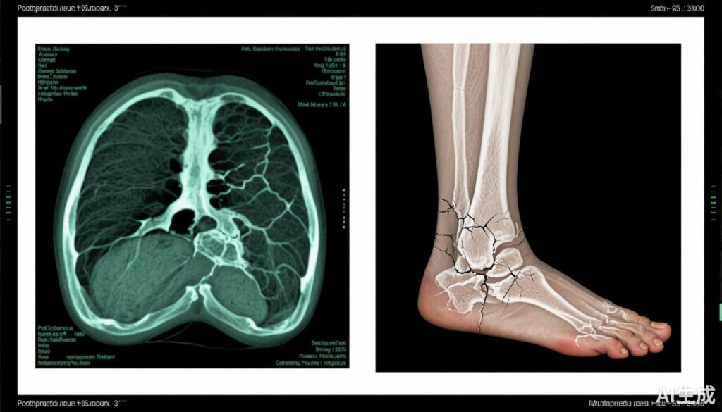
Fracture Characteristics: Tibial fractures were the most frequent, affecting 88% of patients, with foot fractures noted in 49%. Critically, 76% of the cohort presented with multiple stress fractures simultaneously, and 63% experienced recurrent fractures over the follow-up period. Most fractures were atraumatic, implicating methotrexate’s role in bone fragility rather than mechanical trauma.
Imaging Modalities: Initial diagnosis largely relied on plain radiographs (93%), but advanced imaging afforded superior sensitivity and specificity. MRI was performed in 84% of cases, revealing early bone stress injuries and precisely localizing fracture sites. Bone scintigraphy was used in 46%, supporting metabolic activity at fracture sites. PET scans were rarely utilized but may hold complementary diagnostic value.
Treatment and Outcomes: Methotrexate discontinuation was implemented in 77% of patients. Remarkably, 91% of patients who discontinued methotrexate demonstrated favorable clinical and radiographic resolution of fractures, in stark contrast to only 29% in those who continued methotrexate (P < .001). This strong association underscores discontinuation as a critical intervention. Some patients required adjunctive measures for osteoporosis management depending on individual BMD and fracture risk profiles.
Expert Commentary
The study by Robin et al. underscores the need for heightened clinical awareness around methotrexate-induced osteopathy. While MTX remains indispensable for controlling inflammatory arthritis, its potential to contribute to skeletal fragility should not be overlooked, especially in long-term treatment scenarios among postmenopausal women predisposed to bone loss. The pronounced predilection for metaphyseal stress fractures in the tibia and foot aligns with the known microarchitectural bone impairment caused by methotrexate in animal models and limited human data.
Clinically, differentiating methotrexate osteopathy from more common osteoporotic fractures is paramount and challenging. The study supports early use of sensitive imaging modalities such as MRI. Bone scintigraphy serves as a valuable adjunct, particularly when MRI is contraindicated or unavailable. Treatment must include discontinuation of methotrexate whenever methotrexate-induced osteopathy is confirmed, while balancing the risk of arthritis flare-ups.
Limitations of the study include its retrospective design, potential confounding by concurrent corticosteroid use, and the absence of controlled comparison groups. However, the large sample size and multicenter approach add robustness. Future prospective studies are warranted to refine risk stratification, clarify pathophysiological mechanisms, and develop tailored treatment algorithms integrating bone health preservation strategies alongside anti-inflammatory therapies.
Conclusion
Methotrexate-induced osteopathy, though uncommon, represents a clinically significant phenomenon primarily affecting postmenopausal women with RA or PsA on long-term methotrexate therapy. It presents chiefly with atraumatic stress fractures in the tibia and foot, often multiple and recurrent. Early diagnosis facilitated by MRI and bone scintigraphy improves detection, and prompt methotrexate discontinuation markedly enhances clinical outcomes.
Clinicians should maintain vigilance for this condition but also consider that methotrexate’s benefits generally outweigh these risks in most patients. Individualized assessment balancing anti-inflammatory control and bone health is essential. Further research should focus on elucidating underlying mechanisms, identifying predictive markers, and optimizing bone-protective interventions to minimize methotrexate-related skeletal complications.
References
Robin F, Ghossan R, Mehsen-Cetre N, Triquet L, Larid G, Coiffier G, Mina M, Pickering ME, Barthe C, Paccou J, Herman J, Massy E, Roitg I, Branquet M, Lasnier Siron J, Guillouard M, Desmonet Trousset C, Aubrun A, Godfrin B, Hauzeur JP, Chatelus E, Koumakis E, Legrand JL, Schaeverbeke T, Leloix A, Masson M, Nicolau J, Ghiringhelli C, Decrock M, Durel CA, Bouvard B, Cortet B, Casadepax-Soulet C, Malaise O, Javier RM, Briot K, Guggenbuhl P. METHOFRACT, a methotrexate osteopathy multicentre cohort study. RMD Open. 2025 Sep 25;11(3):e005941. doi: 10.1136/rmdopen-2025-005941. PMID: 40998522; PMCID: PMC12481393.


