Patient Information
A 45-year-old female presented to the outpatient dermatology clinic with complaints of recent onset pruritic skin lesions distributed over her trunk and upper limbs. She reported associated symptoms including unexplained weight loss, palpitations, and heat intolerance occurring over a three-month period. Her medical history was significant for no prior thyroid disorders or dermatological conditions. She denied any recent infections or medication changes.
Diagnosis
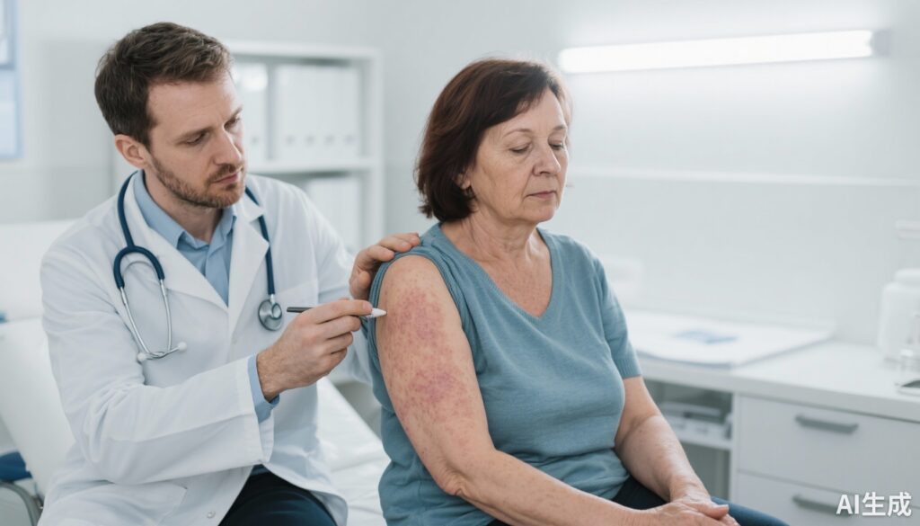
Physical examination revealed warm, moist skin with diffuse erythematous macules and papules accompanied by mild edema over the arms and thorax. Additionally, the patient exhibited fine tremors and mild exophthalmos. Laboratory studies showed elevated serum free T4 and suppressed TSH consistent with hyperthyroidism. A thyroid-stimulating immunoglobulin test was positive, confirming Graves’ disease as the underlying cause. The cutaneous findings were interpreted as manifestations of thyroid dermopathy commonly associated with hyperthyroidism.
Differential Diagnosis
The differential diagnosis for pruritic erythematous skin lesions included:
– Atopic dermatitis: Ruled out due to adult onset without a history of allergy and systemic hypermetabolic signs.
– Psoriasis: Not consistent with lesion morphology or distribution.
– Drug-induced eruptions: No new medications reported.
– Contact dermatitis: No history of exposure to known irritants.
– Other causes of hyperthyroidism-related dermopathy: Thyroid acropachy was not evident.
Overall, the constellation of systemic and cutaneous signs with confirmatory labs supported the diagnosis of hyperthyroidism-associated dermopathy.
Treatment and Management
The patient was initiated on methimazole 15 mg daily to control thyrotoxicosis. Beta-blocker therapy with propranolol was introduced to manage palpitations and tremor. Symptomatic treatment for skin lesions included topical corticosteroids and emollients to alleviate pruritus and inflammation. Patient education about the nature of thyroid dermopathy and the importance of adherence to therapy was provided. Regular monitoring of thyroid function tests was planned every 4–6 weeks.
Outcome and Prognosis
Over a three-month follow-up period, the patient showed significant clinical improvement. Repeat thyroid function tests normalized, and pruritic skin lesions resolved. The patient’s weight stabilized, and symptoms of hypermetabolism abated. Long-term prognosis is favorable with continued treatment adherence. The cutaneous manifestations associated with hyperthyroidism typically resolve with control of thyroid hormone levels.
Discussion
This case illustrates the importance of considering systemic endocrine disorders such as hyperthyroidism in patients presenting with new-onset skin lesions accompanied by systemic symptoms. Graves’ disease, as an autoimmune cause of hyperthyroidism, can present with multiple cutaneous manifestations including dermopathy characterized by pretibial myxedema and less commonly diffuse erythematous pruritic lesions. Recognition of these signs can expedite diagnosis and treatment, reducing morbidity.
Early diagnosis and effective antithyroid treatment are critical for symptom resolution. Beta-blockers provide symptomatic relief until euthyroidism is achieved. Interdisciplinary collaboration between endocrinologists and dermatologists is essential for optimal management. Although cutaneous thyroid dermopathy is generally self-limiting with therapy, persistent or severe cases may require additional interventions such as corticosteroids or immunomodulatory treatments.
This report reinforces established clinical knowledge and highlights the need for comprehensive evaluation of dermatological complaints in the context of systemic disease. Further studies on the prevalence and mechanisms of cutaneous manifestations in hyperthyroid patients could inform improved diagnostic algorithms.
References
1. Smith TJ, Hegedüs L. Graves’ Disease. N Engl J Med. 2016;375(16):1552-1565. doi:10.1056/NEJMra1510030
2. Fatourechi V. Pretibial myxedema: pathophysiology and treatment options. Am J Clin Dermatol. 2005;6(4):295-309.
3. Bahn RS. Graves’ ophthalmopathy. N Engl J Med. 2010;362(8):726-738. doi:10.1056/NEJMra0905750