Highlight
– Baseline radiographic chondrocalcinosis was present in ~5% of osteoarthritis-free knees and predicted subsequent development of radiographic knee osteoarthritis (pooled OR ≈ 1.75).
– Association remained statistically significant among knees with Kellgren-Lawrence grade 0 at baseline (pooled OR ≈ 1.77), supporting a possible etiologic role rather than simple co-occurrence.
– No consistent link was observed between baseline chondrocalcinosis and subsequent incident knee pain, indicating a dissociation between radiographic structural change and symptomatic expression.
Background
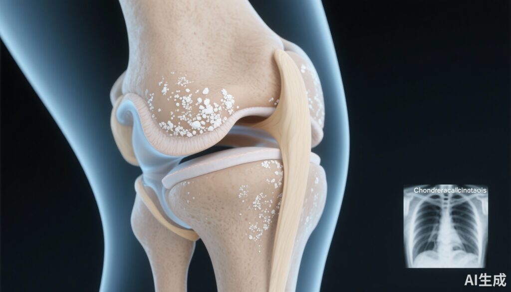
Chondrocalcinosis is the radiographic appearance of calcium-containing crystal deposition in cartilage and fibrocartilage, most commonly reflecting calcium pyrophosphate (CPP) crystal deposition. CPP deposition disease (CPPD) commonly presents as acute inflammatory arthropathy (pseudogout) but is also frequently observed on imaging as chondrocalcinosis in older adults.
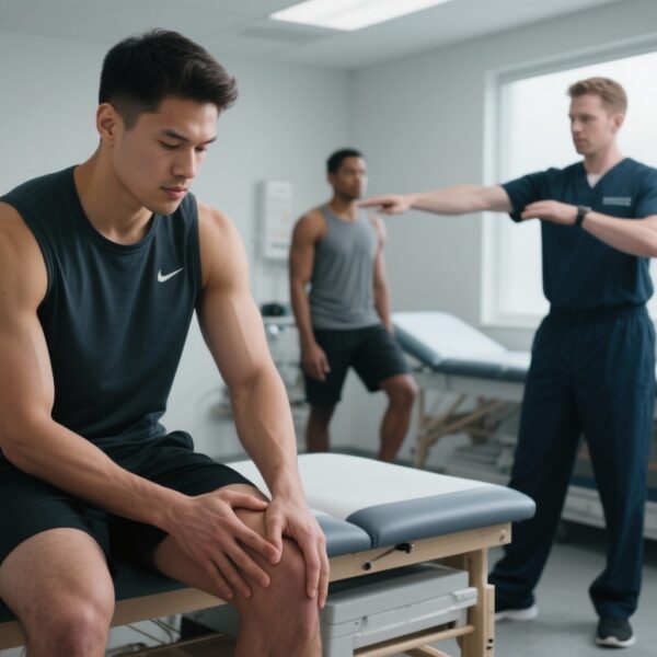
Osteoarthritis (OA) is the most common joint disorder worldwide and a leading cause of pain, disability, and health-care utilization. While OA is classically considered a disease of joint mechanics and cartilage wear, growing evidence implicates metabolic, inflammatory, and crystal-mediated pathways in disease initiation and progression. Distinguishing whether chondrocalcinosis is a consequence of OA-related joint tissue changes or a contributing causal factor has potential implications for risk stratification and targeted prevention.
Study design
This report synthesizes findings from Wu et al., who analyzed data from two large, prospective community-based cohorts to examine whether baseline chondrocalcinosis predicts incident radiographic knee OA and incident knee pain.
Populations and follow-up:
- Rotterdam Study (RS): 3,737 participants with up to 20 years of follow-up.
- Multicenter Osteoarthritis Study (MOST): 2,750 participants with up to 7 years of follow-up.
Inclusion: Participants whose knees were radiographically OA-free or had minimal changes (Kellgren-Lawrence grade [KLG] 0 or 1) at baseline; analyses included a subgroup restricted to KLG = 0 knees.
Exposure: Baseline radiographic chondrocalcinosis (presence of calcification in cartilage/fibrocartilage on knee radiographs).
Outcomes: (1) Incident radiographic knee OA (progression to KLG ≥2) and (2) incident knee pain.
Analytic approach: Logistic regression adjusted for age, sex, and body mass index (BMI) within each cohort; results pooled using meta-analysis techniques. Sensitivity analyses evaluated the KLG = 0 subgroup and assessed associations with incident pain.
Key findings
Prevalence of baseline chondrocalcinosis among knees without OA was low (~5%), consistent with age-related CPPD prevalence in community cohorts.
Association with incident radiographic knee OA
Both cohorts showed a significant association between baseline chondrocalcinosis and development of radiographic knee OA during follow-up. The pooled adjusted odds ratio (OR) was 1.75 (95% CI 1.35–2.27; P < .001), indicating approximately 75% higher odds of incident radiographic OA in knees with chondrocalcinosis versus those without, after controlling for age, sex, and BMI.
Importantly, this association persisted when analysis was confined to knees with KLG = 0 at baseline (pooled OR 1.77, 95% CI 1.04–3.01; P = .035). That the relationship held in truly radiographically normal knees strengthens the interpretation that chondrocalcinosis may precede and potentially contribute to incident OA rather than being purely a bystander of established disease.
Association with incident knee pain
The study did not demonstrate a consistent association between baseline chondrocalcinosis and subsequent development of knee pain. Effect estimates were heterogeneous across cohorts and not uniformly statistically significant. This dissociation suggests that CPP crystal deposition as visualized radiographically may contribute to structural OA development without necessarily producing persistent or predictable symptomatic consequences in the general population.
Effect sizes and clinical interpretation
An OR of ~1.75 is a moderate effect size. Given the relatively low baseline prevalence of chondrocalcinosis in an osteoarthritis-free population (~5%), the population attributable risk would be limited, but for affected individuals these findings indicate materially increased risk of radiographic OA over years to decades. The preservation of the association in KLG = 0 knees supports a potential pathogenic contribution of calcium crystal deposition to structural joint degeneration.
Sensitivity and robustness
The study strengths include large sample sizes, long follow-up in one cohort (up to 20 years), standardized radiographic scoring, and replication across two cohorts with different designs and follow-up durations. Multivariable adjustment for key confounders (age, sex, BMI) was performed, and meta-analytic pooling increased precision.
Expert commentary
Mechanistic plausibility: There is biological plausibility for CPP crystals contributing to joint degeneration. In vitro and synovial studies have shown that CPP crystals can stimulate chondrocytes, synoviocytes, and macrophages to release pro-inflammatory cytokines and matrix-degrading enzymes, promoting cartilage breakdown and osteophyte formation. Thus, crystal deposition could be an active driver of structural change even in the absence of overt acute crystal arthritis.
Clinical implications: These findings support considering chondrocalcinosis as a marker of an OA phenotype where crystal-mediated mechanisms contribute to pathogenesis. For clinicians, detection of chondrocalcinosis in a radiographically normal knee might identify patients at elevated risk for later structural OA. However, routine screening radiographs purely to detect chondrocalcinosis are not indicated based on current evidence, given the low prevalence and the uncertain implication for symptoms and treatment.
Therapeutic implications and research opportunities: If CPP crystals contribute to OA development, therapies that reduce crystal formation, enhance crystal dissolution, or blunt crystal-driven inflammation could be explored as preventive or disease-modifying strategies for this OA subset. Examples might include interventions modifying local pyrophosphate metabolism, inhibitors of crystal formation, or targeted anti-inflammatory strategies during key windows. Randomized trials would be required to test whether intervening on CPP-related pathways modifies OA incidence or progression.
Limitations and generalizability: Several limitations warrant consideration. Radiographic detection of chondrocalcinosis underestimates CPP deposition compared with crystal analysis of synovial fluid or more sensitive imaging modalities (e.g., ultrasound, CT), so misclassification of exposure is possible. Residual confounding by unmeasured factors such as knee injury history, joint alignment, or local biomechanics may influence results. The cohorts primarily included older adults from specific geographic populations; applicability to younger populations or other ethnic groups should be approached cautiously. Lastly, the lack of consistent association with pain highlights the common discordance between radiographic change and symptomatology in OA, complicating decisions about clinical management based on imaging alone.
Conclusion
In two large prospective cohorts, radiographic chondrocalcinosis at baseline was associated with substantially increased odds of developing radiographic knee osteoarthritis during long-term follow-up, including among knees that were radiographically normal at baseline. These results support the concept that calcium crystal deposition can contribute to incident OA and identify a potentially distinct OA subgroup driven in part by crystal-mediated mechanisms.
Clinical practice should not yet change on the basis of this single body of observational data alone, but the findings justify prospective mechanistic studies and trials targeting crystal-related pathways. Future work should use more sensitive imaging and synovial-fluid crystal identification, evaluate molecular mediators linking CPP deposition to cartilage catabolism, and test whether modifying CPP biology prevents OA onset or progression.
Funding and clinicaltrials.gov
Primary study funding sources and trial registrations are reported in the original publication: Wu Y et al., Ann Rheum Dis. 2025 (see References). Readers should consult the original article for detailed funding declarations and cohort-specific support statements.
References
1. Wu Y, Liew JW, Boer JD, Westerland M, LaValley M, Voortman T, Bierma-Zeinstra S, Oei EHG, van Meurs JBJ, Neogi T, Boer CG. Chondrocalcinosis and incident knee osteoarthritis: findings from 2 large prospective cohorts with 20 years of follow-up. Ann Rheum Dis. 2025 Oct;84(10):1743-1751. doi: 10.1016/j.ard.2025.07.009. Epub 2025 Aug 5. PMID: 40764183; PMCID: PMC12380518.
2. Felson DT. Osteoarthritis as a disease of mechanics. Osteoarthritis Cartilage. 2013 Jan;21(1):10-15. doi:10.1016/j.joca.2012.10.008.
AI image prompt for article thumbnail
Realistic medical illustration of an adult human knee in semi-transparent anatomic view showing femur, tibia, menisci, and articular cartilage; white radiopaque speckles representing calcium crystal deposits localized to meniscus and cartilage surfaces; inset small grayscale radiograph in corner showing linear chondrocalcinosis; neutral clinical color palette, high detail, journal cover style.